3D CBCT imaging
Three-dimensional imaging may be used when we need to evaluate anatomy that cannot be fully understood on standard 2D images, such as implant sites, selected airway or surgical questions, roots, bone, or other complex areas.
3D Dental Scans + Digital Imaging · Cedar Park
3D imaging, intraoral scans, and clinical photography help us understand structure, plan treatment, and show you what we see—without using every technology for every patient.
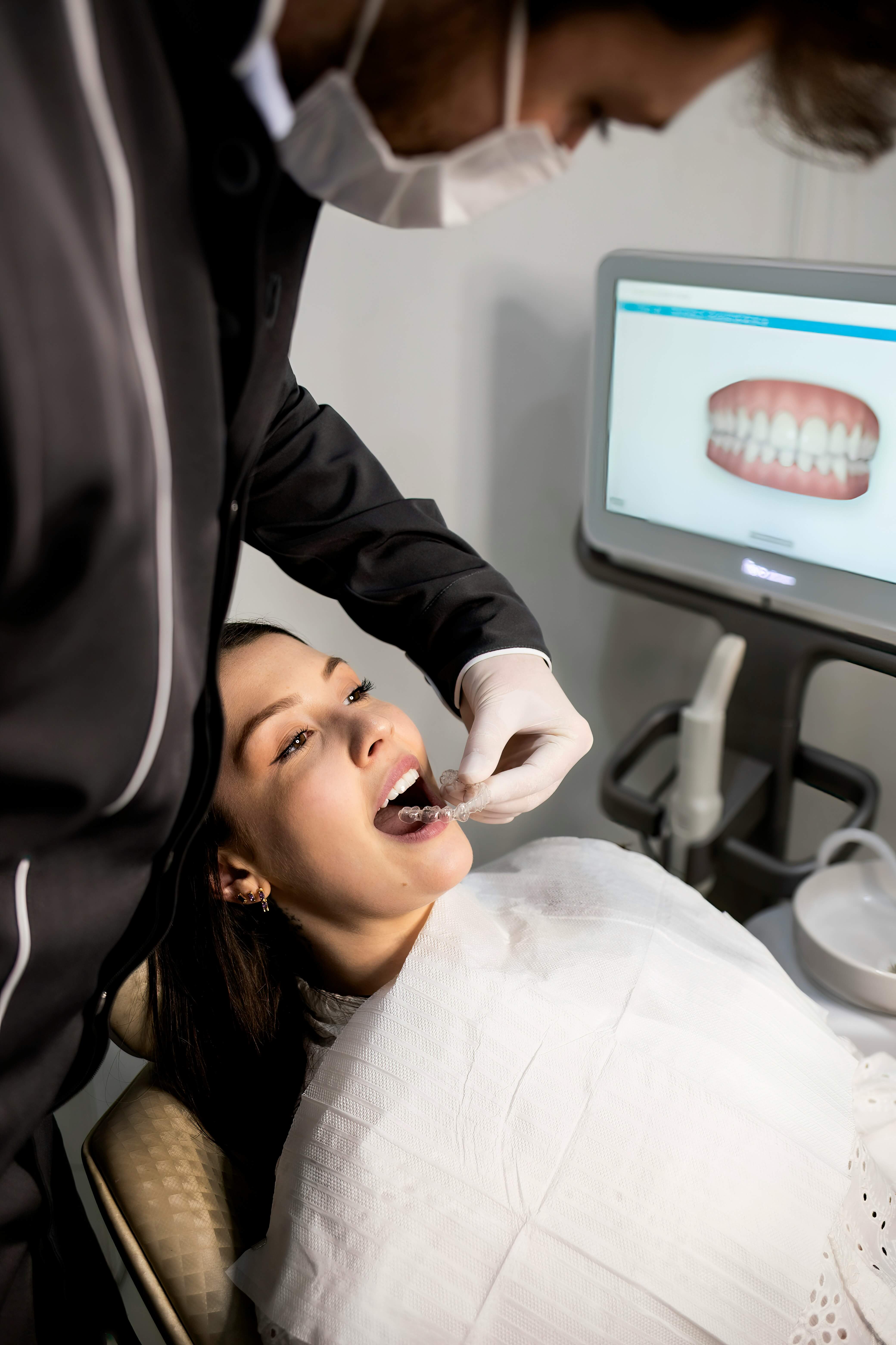
Digital records at Rise
We use different records for different purposes. A routine exam, an implant plan, an airway evaluation, and a cosmetic case do not all require the same imaging.
The goal is to collect the information that actually improves diagnosis, communication, or treatment planning.
What we use
Three-dimensional imaging may be used when we need to evaluate anatomy that cannot be fully understood on standard 2D images, such as implant sites, selected airway or surgical questions, roots, bone, or other complex areas.
A digital scan creates a detailed model of your teeth and bite without traditional impression material and can be used for Invisalign, retainers, restorations, monitoring, and treatment planning.
Photos make it easier to show you what we see, compare changes over time, and plan cosmetic or restorative treatment more clearly.
When digital scans can help
Digital scan FAQ
Cone beam computed tomography creates a three-dimensional view of dental and facial structures. We use it selectively when 3D information is likely to change diagnosis or treatment planning.
No. Standard dental x-rays create two-dimensional images. CBCT provides a three-dimensional dataset and is generally reserved for situations where the additional information is clinically useful.
No. We use 3D imaging selectively based on the clinical question. Routine preventive care often requires different records than implant, surgical, orthodontic, or airway planning.
An intraoral scanner captures the shape of your teeth and bite digitally. It can replace traditional impressions for many procedures and is useful for planning, monitoring, and fabrication.
Photography helps document findings, communicate clearly, track changes, and plan treatment—especially when appearance, wear, alignment, or restorative changes are involved.
Usually these records are selected as part of your exam or treatment planning based on what is needed. You do not need to choose a scan before we evaluate you.